eISSN: 2093-8462 http://jesk.or.kr
Open Access, Peer-reviewed
eISSN: 2093-8462 http://jesk.or.kr
Open Access, Peer-reviewed
Ji-Woo Seok
, Jin-Hun Sohn
10.5143/JESK.2019.38.5.361 Epub 2019 October 31
Abstract
Objective: The purpose of this study is to find brain areas shown a difference of the volume and cortical thickness between females with major depressive disorder (MDD) and healthy females.
Background: Although neuroimaging studies on the characteristics of MDD have been accumulating, limited studies have used both brain volume and cortical thickness to find the structural alteration in MDD. Integrating measures of structural brain imaging provide profound insights into the neurobiological underpinnings of MDD.
Method: Twenty-two females with MDD and 23 age- and sex-matched healthy females participated in this study. A 3T Philips Achieva MRI scanner (Philips Medical Systems, Best, The Netherlands) was used for image acquisition. Gray matter volume and cortical thickness were analyzed using voxel-based morphometry (VBM) and surface-based morphometry (SBM).
Results: VBM results indicated that individuals with MDD had significantly reduced gray matter volume in the anterior cingulate gyrus, hippocampus, hippocampal gyrus and precuneus and cerebellum compared to controls. Moreover, gray matter volume in the right anterior cingulate gyrus was negatively correlated with the severity of depression. SBM results demonstrated that compared to healthy controls, individuals with MDD showed reduced cortical thickness in temporal pole, paracentral gyrus, angular gyrus and insula.
Conclusion: These findings suggest that structural alterations in the anterior cingulate gyrus are linked to MDD, thereby providing understanding into the neural mechanisms underlying MDD.
Application: Alterations in the anterior cingulate cortex may act as neuroimaging markers of MDD, and these findings provide a possibility to developed the diagnosis system for MDD.
Keywords
Major depressive disorder Voxel-based morphometry Surface-based morphometry Anterior cingulate cortex
주요우울장애는 전 세계적으로 평생 유병률이 5~15%에 해당하는 가장 흔한 정신질환으로 무쾌감증과 함께 인지 및 사회적 기능 장애를 동반한다(Kessler, 2003). 뇌 영상 분석 기법의 발달에 따라 뇌내 신경회로의 구조적, 기능적 변화가 주요우울장애의 기분 저하, 인지 및 사회적 기능 장애에서 중요한 역할을 할 것이라고 제안되고 있다(Busatto, 2013). 즉, 정서 및 인지 기능과 관련된 피질-변연 회로 내의 뇌 영역들의 활성도의 저하와 네트워크내 연결성 저하가 주요우울장애의 기능 장애와 관련이 있다고 보고되고 있다(Kaiser et al., 2015; Pizzagalli, 2011; Tu et al., 2012).
최근에는 주요우울장애 환자들의 뇌의 기능적인 측면뿐만 아니라 뇌의 부피 및 피질 두께를 이용하여 뇌의 구조적 변화를 분석한 연구들도 보고되고 있다(Bora et al., 2012; Koolschijn et al., 2009; Lai, 2013).
주요우울장애 환자의 국소적인 회백질 위축에 대한 연구들은 전전두엽(Kumar et al., 1998), 전측 대상 피질(Caetano et al., 2006), 안와전두 피질(Monkul et al., 2007), 그리고 해마(Maller et al., 2007)의 부피 감소를 보고하였다. 이 연구들은 뇌의 전체 영역 혹은 특정 영역을 관심 뇌 영역(Region of Interest, ROI)으로 지정하고, 자기공명영상을 바탕으로 전문가가 관심 뇌 영역을 직접 그려서 해당부분의 부피를 측정하는 방법을 사용하였다. 이러한 전통적인 형태계측법은 시간 소모적이며, 연구자의 주관적 판단에 의존할 수밖에 없어 오차가 크다는 단점이 제기되어 왔다.
뇌의 부피 변화를 측정하기 위하여 사용되는 복셀기반 형태계측(voxel-based morphometry, VBM) 분석법은 두뇌 전체를 복셀 단위로 쪼개어 국소적인 뇌의 변형을 통계적으로 측정함으로써 회백질과 백질의 밀도 또는 농도와 함께 부피의 차이를 관찰하는 방법이다(Ashburner and Friston, 2000). 복셀기반 형태계측 기술은 뇌의 전체적인 변화를 객관적이고 자동적으로 조사할 수 있다는 장점을 가지고 있어, 우울증의 회백질 부피 변화를 측정하기 위해 자주 사용되는 추세이다(Bora et al., 2012; Koolschijn et al., 2009; Lai, 2013). 복셀기반 형태계측법을 사용한 23개의 연구로부터 수집된 데이터를 이용한 메타분석 연구에 따르면, 주요우울장애 환자의 전측 대상 피질의 회백질의 부피와, 배측 전전두 피질의 회백질의 부피가 감소했다(Bora et al., 2012). 또다른 20개의 복셀기반 형태계측 연구에 대한 메타분석 결과도 주요우울환자가 정상군과 비교했을 때 양측 전측 대상 피질의 회백질의 부피 저하가 보고되었다(Lai, 2013).
최근에 개발된 표면기반 형태계측(surface-based morphometry, SBM) 분석법은 뇌의 이랑과 고랑의 패턴에 따른 정합을 하여 뇌 이랑과 고랑의 해부학적 변이를 정확하게 측정할 수 있을 뿐 아니라, 뇌 회백질의 두께를 측정한다는 장점이 있다(Dahnke et al., 2013). 형태계측 분석법을 이용한 연구에서, 정상군과 비교했을 때 주요우울 환자들은 전두엽과 하두정엽에서 더 얇은 피질 두께를 보였다(Tu et al., 2012). 이렇듯 다양한 형태계측법을 이용하여 주요우울 환자 내 뇌의 구조적 변화를 측정하려는 시도가 이루어지고 있으나, 다양한 형태계측 방법을 결합하여, 뇌의 용적과 피질의 두께 분석을 동시에 수행한 우울증 뇌 영상 연구는 거의 이루어지지 않고 있는 실정이다.
따라서 본 연구에서는 결과의 교차타당도를 높이고 주요우울장애 특징적인 뇌 내 구조물을 규명하기 위하여, 다양한 형태계측 방법을 적용하고자 한다. 즉 주요우울 환자군과 정상군에서 복셀기반 형태계측법과 표면기반 형태계측법을 통해 용적 측정과 피질 두께 측정을 시행하고, 정상군과의 비교를 통해 의미 있는 차이를 보이는 뇌 내 구조물을 규명할 것이다.
2.1 Participants
22명의 주요우울장애 환자 집단(여성, 평균나이 21.50, 표준편차 2.46)이 뇌 영상 실험에 참여하였다. 나이와 성별, 그리고 향정신성 약물 복용 경험에 따라 대뇌 부피 변화와 관련이 있다는 선행 연구에 따라, 본 연구에서 주요우울장애 환자군의 포함기준은 정신장애 진단 및 통계 편람 제 4판에서 주요우울장애 진단기준을 충족하는 20세에서 30세 사이의 향정신성 약물의 복용 경험이 전혀 없는 여성 환자만으로 정하였다. 주요우울장애 진단은 임상전문가에 의해 구조적 면담에 의한 진단 도구인 Structured Clinical Interview for DSM-IV Axis I disorders (SCID-1)를 이용하여 진단되었다. 정상군의 경우 우울 환자군과 나이, 학력 등 인구통계학적 특성이 유사하고, 현재 또는 과거에 Axis I 혹은 II 진단을 받은 적이 없는 여성 23명(여성, 평균나이 21.48, 표준편차 2.66)을 모집하였다.
본 연구는 충남대학교의 생명윤리위원회에 승인(201309-SB-004-01)을 받았으며, 모든 참가자는 휴지기 상태의 기능적 자기공명영상 촬영에 대한 목적과 절차에 대해 충분한 설명을 들은 후 서면으로 동의하였다.
2.2 Assessments
모든 피험자에서 우울증상의 심각도와 임상적 특성을 평가하기 위해 Beck의 우울 척도(Beck Depression Inventory, BDI), Hamilton 우울 평가 척도(Hamilton Depression Rating Scale, HDRS), 자살사고 질문지(Suicidal Ideation Questionnaire, SIQ), Beck의 불안 척도(Beck Anxiety Inventory, BAI), 지각된 스트레스 척도(Perceived Stress Scale, PSS)가 사용되었다.
Beck의 BDI 척도는 정서적, 인지적, 동기적 및 생리적 증상 영역을 측정하며 우울 증상의 심각도를 평가하는 자기 보고형 척도로 총 21개의 문항으로 구성되어 있다. 점수의 범위는 0점에서 63점까지이며, Cronbach's alpha의 값은 .89였다(Hahn, 1982). Hamilton의 우울 평가 척도는 관찰자형 평가 척도로 임상적 판단을 수치화하기 위하여 개발된 척도이다. 총 17문항으로 구성되어 있으며, 총점은 0점에서 52점까지로, Cronbach's alpha의 값은 .76 이였다(Hamilton, 1986; Lee et al., 2005). 자살사고를 측정하는 도구는 Reynolds (1988)에 의해 개발된 자살사고 질문지는 현재의 자살생각의 수준을 측정하는 문항으로 이루어진 자기 보고식 척도이다. 총 30문항으로 Cronbach's alpha의 값은 .93이였다(Reynolds and Mazza, 1999). Beck 등이 개발한 불안 척도(Beck et al., 1988)는 한 주 동안 개인이 경험한 불안 증상의 정도를 4점 척도 상에서 평정하도록 하는 자기 보고식 설문지로 21개의 문항으로 구성되어 있다. 총점은 0~63점까지이며, Cronbach's alpha의 값은 .89이였다. Cohen et al. (1983)이 개발한 지각된 스트레스 척도는, 지난 한 달 동안 개인의 생활이 예측할 수 없고, 조절할 수 없으며, 부담이 되었다고 지각하는 정도를 의미한다. 자기 보고식 설문지로 10개 항목으로 구성되어 있으며, 점수범위는 0에서 40점까지이다. Cronbach's alpha는 .78이었다.
2.3 Imaging parameter
뇌 영상 실험은 한국기초과학지원연구원에 있는 MRI scanner (3.0T Philips MR scanner)를 사용하였다. 파라미터의 조건은 TR/TE=280 /14ms, flip angle = 60°; field of view (FOV) = 24 × 24cm2; matrix = 256 × 256; slice thickness = 4mm without gap, 35 slices였다. 뇌 영상 촬영은 약 5분간 진행되었으며, 피험자에게 뇌 영상 촬영을 하는 동안 눈은 감은 상태에서 최대한 머리를 움직이지 말라고 지시하였다.
2.4 Analysis for Voxel-based morphometry method
T1 영상 자료는 SPM12 (Statistical Parametric Mapping; Wellcome Department of Cognitive Neurology, London, UK)와 DARTEL toolbox를 이용하여 분석하였다. 각 개인의 영상과 표준공간의 정합도(coregistration accuracy)를 높이기 위해 전교련(anterior commissure)이 영상의 중심 좌표가 되도록 조정한 후, 바이어스필드에 의해 왜곡된 뇌 영상 자료(bias-field inhomogeneities)를 비모수 비균일 강도 표준화(non-parametric non-uniform intensity normalization)를 통해 신호를 보정하였다. 이 후, 각 개인의 영상을 회백질, 백질, 뇌실로 분할(segmentation)하고, 피험자간 동질성(inter-subject homogeneity)과 전체적인 영상의 질을 검증하기 위하여 분할 후 동질성 검사(check homogeneity)를 실시하여 변산성이 큰 영상은 분석에서 제외하였다. 해부학적 정확성을 향상시키기 위해 DARTEL 알고리즘을 적용하여 공간적으로 정규화(normalization) 시켰다(Ashburner, 2007). 정규화된 뇌 영상들은 MNI 공간(MNI space)에 따라 워핑(warping)되었고, 워핑에 의해 발생할 수 있는 볼류 효과를 보정하기 위해 야코비안 행렬(Jacobian determinant)을 이용함으로서 각 화소에 적용되는 회백질의 양을 보존하였다. 마지막으로 8mm 가우시안 커널(gaussian kernel)을 이용하여 평편화(smoothing) 되었다. 전처리 과정을 거친 최종 회백질의 영상의 각 복셀(voxel)의 크기는 2×2×2였으며, 각 복셀의 신호값은 회백질의 부피 정보를 반영한다.
2.5 Analysis for Surface-based morphometry method
표면기반 계측법의 경우 SPM12 (Statistical Parametric Mapping; Wellcome Department of Cognitive Neurology, London, UK)에서 구현된 CAT 12 toolbox의 파이프라인을 이용하여 분석하였다. 각 개인별로 영상의 중심 좌표를 재조정한 후 비모수 비균일 강도 표준화(non-parametric non-uniform intensity normalization)를 통해 신호를 보정하였다. 이후 정확도를 높이기 위하여 부분 부피 보정(partial volume correction)과 고랑 흐림과 고랑 비대칭성의 보정(correction for sulcal blurring and sulcal asymmetries)을 적용한 투영기반 두께 추정법(projection-based thickness estimation)을 이용하여, 피질 두께(cortical thickness)를 계산하였다. 또한 구면조화함수(spherical harmonics)를 적용하여 위상 보정(Topological correction)이 수행되었다. 표면의 재매개화(reparameterization)를 위해, 감소한 국소왜곡이 나타난 피질 표면의 구형 매핑을 위한 알고리즘이 적용되었다. 표면의 구형 정합을 위한 방법으로는 2차원 DARTEL 알고리즘이 사용되었다(Yotter et al., 2011). 이랑화 지수(gyrification index)는 절대 평균 곡률 접근법(absolute mean curvature approach)에 기초하여 추출되었다(Luders et al., 2006). 마지막으로, 모든 표면 측정은 15mm (FWHM)의 가우시안 커널(gaussian kernel)로 다시 샘플링하고 편평화 했다.
2.6 Statistical analysis for behavioral data
우울 증상의 심각도와 임상적 특성은 독립 표본 t 검정을 이용하여 집단간 비교를 하였다. 복셀기반 형태계측법과 표면기반 형태계측법을 통해 얻어진 자료들로부터 주요우울장애군과 정상군 간의 회백질 부피 및 피질 두께 차이를 SPM 8 내의 독립 표본 t 검정(p<0.001, uncorrected)으로 분석하였으며, 20복셀 이하의 작은 크기의 클러스터들은 제외되었다.
3.1 Demographic and psychological characteristics
본 연구의 대상자들에 대한 인구학적, 임상적 특성은 다음과 같다(Table 1). 주요우울장애군과 정상군 사이에서 연령에서는 유의미한 차이를 보이지 않았으나(p>0.05), Beck의 우울척도 점수와 Hamilton의 우울평가 척도 점수, 자살사고 질문지 점수, Beck의 불안척도 점수, 지각된 스트레스 점수에서는 유의미한 차이가 나타났다.
|
|
MDD |
Controls |
t value |
p value |
|
Age |
21.50
(2.46) |
21.48
(2.66) |
0.03 |
0.97 |
|
BDI |
33.73
(7.87) |
5.52
(3.29) |
15.81 |
< .001 |
|
HDRS |
20.36
(6.63) |
2.87
(2.32) |
11.92 |
< .001 |
|
SIQ |
71.14
(39.69) |
5.74
(6.23) |
7.81 |
< .001 |
|
BAI |
23.36
(13.69) |
3.74
(3.86) |
6.61 |
< .001 |
|
PSS |
18.63
(5.13) |
14.48
(5.36) |
10.66 |
.011 |
3.2 Brain volume difference between individuals with major depressive disorder and healthy controls
전체 뇌 용적의 경우 주요우울장애군은 1477±197mL, 정상군은 1444±170mL으로 유의미한 차이를 보이지 않았다(t=0.62). 뇌 회색질의 용적은 주요우울장애군의 경우 671±66mL, 정상군의 경우 671±75mL으로 유의미한 차이가 나타나지 않았다(t=.0049). 뇌 척수액(t=0.76)과 뇌 백색질(t=0.74)의 경우에도 두 집단간 차이가 나타나지 않았다.
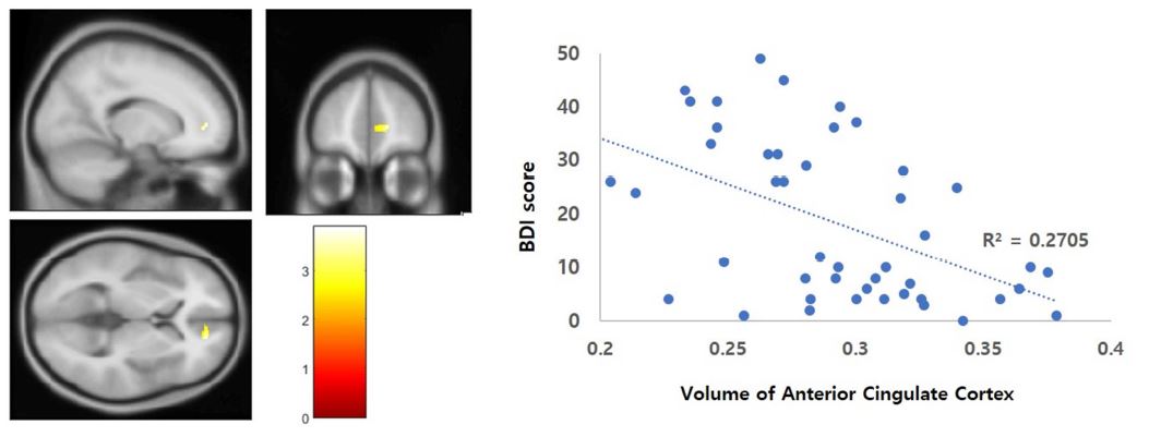
우측 대상회(right anterior cingulate gyrus), 우측 쐐기앞소엽, 좌측 해마(left hippocampus)와 해마방회(left parahippocampal gyrus), 좌측 소뇌에서 주요우울장애군이 정상군에 비해 유의한 부피 감소를 보였다(p<0.001, uncorrected) (Table 2, Figure 1). 주요우울장애군에 비해 정상군이 부피감소를 보이는 뇌 영역은 없었다.
|
Brain areas |
MNI coordinates |
t values |
Voxel size |
||||||||||
|
x |
y |
z |
|||||||||||
|
MDD < Controls |
|||||||||||||
|
Right anterior cingulate cortex
(ACC) |
14 |
45 |
-2 |
3.95 |
391 |
||||||||
|
Right precuneus |
6 |
-51 |
55 |
3.66 |
186 |
||||||||
|
Left hippocampus /
parahippocampal gyrus |
-35 |
-27 |
-18 |
3.39 |
127 |
||||||||
|
Left cerebellum |
17 |
-38 |
-51 |
3.30 |
255 |
||||||||
|
Result of correlation analysis |
|||||||||||||
|
Right anterior cingulate cortex (ACC) |
17 |
50 |
3 |
3.35 |
113 |
||||||||

주요우울장애군에서 Beck의 우울척도 점수로 계산된 우울증 심각도와 대상회(anterior cingulate gyrus) 영역의 뇌 회색질 부피와 부적 상관관계(r=-0.52, p<0.001)가 있는 것으로 나타났으며, 다른 임상검사 결과와 통계적으로 유의한 상관관계를 보이는 영역은 없었다(Table 2, Figure 2).
3.3 Brain surface difference between individuals with major depressive disorder and healthy controls
전체 뇌 피질 두께의 경우 주요우울장애군은 2.58±0.10mm, 정상군은 2.61±0.06mm으로 유의미한 차이를 보이지 않았다(t=1.19).
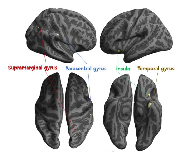
한편 좌측 측두엽(left temporal cortex), 우측 상변연회(right angular gyrus), 우측 중심방소엽(right precuneus), 좌측 뇌섬엽(right insula)에서 주요우울장애군은 정상군에 비해 유의미하게 얇은 피질 두께를 보였다(Table 3, Figure 3).
주요우울장애군에서 우울증 심각도나 다른 임상검사 결과와 유의미한 상관관계를 보이는 영역은 없었다.
|
Brain areas |
MNI coordinates |
t value |
Voxel size |
||
|
x |
y |
z |
|||
|
MDD < controls |
|||||
|
Left temporal gyrus |
-31 |
3 |
-43 |
3.98 |
72 |
|
Right supramarginal gyrus |
54 |
-27 |
40 |
3.75 |
16 |
|
Right paracentral gyrus |
16 |
-44 |
53 |
3.49 |
18 |
|
Left insula |
-28 |
21 |
-13 |
3.47 |
22 |
본 연구는 복셀기반 형태분석법과 표면기반 형태분석법을 적용하여 약물을 복용하지 않는 주요우울장애 여성 환자군의 뇌의 구조적 이상을 탐색하고자 하였다. 본 연구 결과, 정상군과 비교하였을 때, 주요우울장애 환자군은 좌측 측두엽과 좌측 뇌섬엽, 우측 상변연회, 우측 중심방소엽의 피질 두께가 얇아지는 것을 관찰하였다. 또한, 피질 두께의 경우, 정상 대조군과 비교하였을 때 주요우울장애 환자군의 경우 우측 대상회, 우측 쐐기앞소엽, 좌측 해마 및 해마방회와 좌측 소뇌 영역에서 유의미하게 부피가 작아짐을 관찰하였다. 특히, 우측 대상회의 경우 우울증의 심각도에 따라 부피가 감소함이 발견되었다.
본 연구의 피질 두께 분석 결과, 주요우울장애군에서 측두엽의 피질 두께 감소를 관찰하였다. 측두엽은 정서 조절 및 사회인지와 관련된 영역으로 알려져 있으며, 주요우울장애 환자의 경우 이 영역의 회백질 감소와 이 영역을 포함한 뇌 회로의 기능적 연결성 저하가 관찰된 바 있다(Cullen et al., 2009; Shah et al., 2002; Takahashi et al., 2010). Takahashi 등 (2010)는 측두엽의 용적 감소가 약물 사용, 증상의 호전과 상관없이 지속적으로 나타남을 밝힘으로써, 측두엽의 부위가 주요우울장애의 발생 및 재발과 관련되는 기질적인 특성일 수 있다고 제안하였다.
또한 정상군에 비해 주요우울장애 환자군에서 소뇌의 부피 감소가 나타났다. 주요우울장애와 관련하여 많은 뇌 구조 영상 연구가 있어 왔지만, 주로 대뇌 피질의 변화에만 주목하여 소뇌의 형태학적 변화를 보고한 연구는 드문 편이다. 하지만 최근에 주요우울장애 환자군에서 소뇌의 회백질 밀도 감소 등이 보고되고 있는 추세이다(Frodl et al., 2008; Peng et al., 2011). 또한 Depping et al. (2016)은 주요우울장애 환자 중 증상이 완화된 환자에서 소뇌 소엽 IX 영역의 부피가 유의미하게 커졌음을 보고하였다. 이 결과는 소뇌가 뇌가 손상을 받을 취약성과 증상을 완화시키는 뇌의 회복력의 기전과 관련되어 있을 수 있음을 시사한다(Depping et al., 2016).
측두엽 이외에도 뇌섬엽의 피질 두께가 얇아짐이 나타났는데, 주요우울장애 환자를 대상으로 한 연구에서 뇌섬엽의 활동성 저하와 대사 이상, 그리고 기능적 연결성 이상은 꾸준히 보고되어 왔다(Avery et al., 2014; Horn et al., 2010; Wiebking et al., 2010). 이들에 따르면, 뇌섬엽은 내적자각(interoceptive awareness)과 관련된 영역으로 주요우울장애 환자의 주요 특징인 여러 가지 통증이나 불편감을 호소하는 신체 증상이 이와 관련 있다고 제안하였다. 즉 뇌섬엽의 기능 이상으로 인해 내적자각 능력이 저하되고, 이로인해 우울증을 통증과 같은 신체 증상으로 오인할 수 있다고 하였다. 또한 뇌섬엽은 대상회와 긴밀한 해부학적 연결성을 가지고 있으며, 대상회와 함께 주의관련 회로(attention network)를 구성하고 있다.
대상회의 경우 많은 메타 연구에서 밝혀졌듯 주요우울장애에 가장 핵심적인 뇌 영역으로 알려져 있으며(Bora et al., 2012; Lai, 2013), 본 연구 결과에서도 주요우울장애군에게 대상회의 부피 감소가 발견되었다. 특히 대상회의 부피 감소가 우울증의 심각도와 유의한 상관을 보여 이 영역의 이상이 우울증의 핵심적인 위험인자로 작용할 수 있음을 의미한다. 대상회는 주의집중능력 및 실행 기능과 관련된 영역으로, 주요우울장애 환자의 임상적 특징으로 나타난 주의집중 곤란, 정신운동 지체, 그리고 저하된 인지 능력 등이 이 영역의 이상과 연관이 있는 것으로 추정된다(Demyttenaere et al., 2005).
대상회 이외에도, 본 연구 결과에서 관찰된 해마 및 해마방회, 두정엽 등은 주요우울장애 환자군에서 보이는 인지적 기능 저하와 관련이 있을 것으로 보인다. 해마의 경우 주요우울장애의 메타분석 연구에서 부피 감소가 일관되게 관찰되었으며(McKinnon et al., 2009), 주요우울장애 환자의 자녀들도 정상군에 비해 부피 감소가 보고되었다(Chen et al., 2010). 또한 주요우울장애 환자에서 두정엽의 부피 감소가 종종 보고되고 있으며(Ries et al., 2009), 본 연구와 유사하게 약물 복용력이 없는 젊은 환자만을 대상으로 한 형태계측 연구에서도 해마와 두정엽의 구조 이상이 보고되었다(Zou et al., 2010). 과제기반 기능적 자기공명영상 연구에 따르면, 해마는 명시적 기억(implicit memory)과 두정엽은 주의 전환(attention shifting)와 관련된 영역으로 알려져 있다(Osaka et al., 2007; Ranganath et al., 2005; Tamm et al., 2004). 이 연구들에서, 실행 기능의 저하와, 기억력의 저하를 보이는 피험자들은 두정엽과 해마의 활동이 감소하였음이 보고되었다. 이에따라 본 연구 결과에서 보고된 주요우울환자군의 대상회, 해마 및 해마방회, 두정엽의 구조적 이상은 우울 증상의 주된 특징인 인지 기능 저하 즉, 정신운동 지체, 집중력 저하, 주의 전환, 의사결정 능력의 저하, 서술적 기억 및 명시적 기억 능력의 감소가 나타난다는 사실에 부합하는 결과이다.
종합해보면, 본 연구의 결과에서 나타난 대상회와 뇌섬엽, 측두엽의 구조적 이상은 전전두엽-변연계 회로의 변화가 주요우울장애의 병태생리에 중요한 역할을 한다는 기존의 연구 결과를 지지할 뿐 아니라, 두정엽, 해마 및 해마방회의 구조적 이상을 밝혀 주요우울장애의 임상적 특징인 인지 기능 저하에 대한 병인학적 증거를 제공하였다.
본 연구의 제한점은 다음과 같다. 첫째, 종단 연구가 아닌 횡단 연구로 진행되었기 때문에 본 연구에서 나타난 뇌 영역의 구조 변화와 우울증의 인과관계를 설명하는 데는 한계가 있다는 점이다. 둘째, 본 연구에서는 여성 우울장애 환자군만을 대상으로 하였기 때문에, 본 연구 결과가 남성 우울장애 환자군에서도 같은 결과가 나올지는 미지수이다. 따라서 본 연구 결과를 우울장애 환자군 모두에게 적용시키는 것에는 한계가 있다. 이니 추후연구를 통해 우울장애를 가진 남성환자군에서도 동일한 뇌 영역에 구조적 이상이 발견되는지 밝혀질 수 있을 것이다. 마지막으로, 형태계측 분석 시 뇌의 크기에 대한 개인차를 보정하기 위하여 전체 뇌 체적을 통제하였음에도 불구하고, 본 연구 결과에서 개인차에 기인한 결과일 가능성을 완전히 배제할 수는 없다. 왜냐하면 뇌의 부피와 피질 두께의 경우, 선천적으로나 후천적인 발달과정상의 문제로부터 영향을 받을 수 있기 때문이다.
하지만 이러한 한계점에도 불구하고, 본 연구는 뇌의 구조적 변화에 영향을 미칠 수 있는 동반질환과 약물 복용이력, 성별 등의 변인들을 통제함으로써 우울장애 특징적인 병인학적 요소에 대한 정보를 제공하였다. 또한 뇌의 구조적 이상을 부피와 피질의 두께 등 다각적인 차원에서 입증함으로써 본 연구 결과의 타당성을 입증했다는데 의의가 있다.
References
1. Ashburner, J. and Friston, K.J., Voxel-based morphometry—the methods. Neuroimage, 11(6), 805-821, 2000.
Google Scholar
PubMed
2. Ashburner, J., A fast diffeomorphic image registration algorithm. Neuroimage, 38(1), 95-113, 2007.
Crossref
Google Scholar
PubMed
3. Avery, J.A., Drevets, W.C., Moseman, S.E., Bodurka, J., Barcalow, J.C. and Simmons, W.K., Major depressive disorder is associated with abnormal interoceptive activity and functional connectivity in the insula. Biological Psychiatry, 76(3), 258-266, 2014.
Crossref
Google Scholar
4. Beck, A.T., Epstein, N., Brown, G. and Steer, R.A., An inventory for measuring clinical anxiety: psychometric properties. Journal of Consulting and Clinical Psychology, 56(6), 893, 1988.
Crossref
Google Scholar
5. Bora, E., Fornito, A., Pantelis, C. and Yücel, M., Gray matter abnormalities in major depressive disorder: a meta-analysis of voxel-based morphometry studies. Journal of Affective Disorders, 138(1-2), 9-18, 2012.
Google Scholar
6. Busatto, G.F., Structural and functional neuroimaging studies in major depressive disorder with psychotic features: a critical review. Schizophrenia Bulletin, 39(4), 776-786, 2013.
Google Scholar
7. Caetano, S.C., Kaur, S., Brambilla, P., Nicoletti, M., Hatch, J.P., Sassi, R.B., Mallinger, A., Matcheri, S., Keshavan, S., David, J., Ellen, F. and Soares, J.C., Smaller cingulate volumes in unipolar depressed patients. Biological Psychiatry, 59(8), 702-706, 2006.
Crossref
Google Scholar
8. Chen, M.C., Hamilton, J.P. and Gotlib, I.H., Decreased hippocampal volume in healthy girls at risk of depression. Archives of General Psychiatry, 67(3), 270-276, 2010.
Crossref
Google Scholar
9. Cohen, S., Karmarck, T. and Mermelstein, R., A global measure of perceived stress. Journal of health and Social Behavior, 24(4), 385-396, 1983.
Google Scholar
10. Cullen, K.R., Gee, D.G., Klimes-Dougan, B., Gabbay, V., Hulvershorn, L., Mueller, B.A., Camchong, J., Bell, J.B., Houri, A., Kumra, M.D., Castellanos, F.X., Lim, K.O. and Milham, M.P., A preliminary study of functional connectivity in comorbid adolescent depression. Neuroscience Letters, 460(3), 227-231, 2009.
Google Scholar
11. Dahnke, R., Yotter, R.A. and Gaser, C., Cortical thickness and central surface estimation. Neuroimage, 65, 336-348, 2013.
Google Scholar
12. Demyttenaere, K., De Fruyt, J. and Stahl, S.M., The many faces of fatigue in major depressive disorder. International Journal of Neuropsychopharmacology, 8(1), 93-105, 2005.
Google Scholar
13. Depping, M.S., Wolf, N.D., Vasic, N., Sambataro, F., Hirjak, D., Thomann, P.A. and Wolf, R.C., Abnormal cerebellar volume in acute and remitted major depression. Progress in Neuro-Psychopharmacology and Biological Psychiatry, 71, 97-102, 2016.
Google Scholar
14. Frodl, T.S., Koutsouleris, N., Bottlender, R., Born, C., Jäger, M., Scupin, I., Reiser, M., Moller, H.J. and Meisenzahl, E.M., Depression-related variation in brain morphology over 3 years: effects of stress?. Archives of General Psychiatry, 65(10), 1156-1165, 2008.
Google Scholar
15. Hahn, H.M.Y.T., A standardization study of Beck Depression Inventory in Korea. Journal of Korean Neuropsychiatric Association, 25, 487-502, 1982.
Google Scholar
16. Hamilton, M., The Hamilton rating scale for depression. In Assessment of depression (pp. 143-152). Springer, Berlin, Heidelberg, 1986.
Crossref
Google Scholar
17. Horn, D.I., Yu, C., Steiner, J., Buchmann, J., Kaufmann, J., Osoba, A., Eckert, U., Ziehut, K.C., He, H., Biswal, B., Bogert, B. and Walter, M., Glutamatergic and resting-state functional connectivity correlates of severity in major depression-the role of pregenual anterior cingulate cortex and anterior insula. Frontiers in Systems Neuroscience, 4, 33, 2010.
Google Scholar
18. Kaiser, R.H., Andrews-Hanna, J.R., Wager, T.D. and Pizzagalli, D.A., Large-scale network dysfunction in major depressive disorder: a meta-analysis of resting-state functional connectivity. JAMA Psychiatry, 72(6), 603-611, 2015.
Crossref
Google Scholar
19. Kessler, R.C., Epidemiology of women and depression. Journal of Affective Disorders, 74(1), 5-13, 2003.
Crossref
Google Scholar
PubMed
20. Koolschijn, P.C.M., van Haren, N.E., Lensvelt‐Mulders, G.J., Hulshoff Pol, H.E. and Kahn, R.S., Brain volume abnormalities in major depressive disorder: A meta‐analysis of magnetic resonance imaging studies. Human Brain Mapping, 30(11), 3719-3735, 2009.
Google Scholar
21. Kumar, A., Jin, Z., Bilker, W., Udupa, J. and Gottlieb, G., Late-onset minor and major depression: early evidence for common neuroanatomical substrates detected by using MRI. Proceedings of the National Academy of Sciences, 95(13), 7654-7658, 1998.
Google Scholar
22. Lai, C.H., Gray matter volume in major depressive disorder: a meta-analysis of voxel-based morphometry studies. Psychiatry Research: Neuroimaging, 211(1), 37-46, 2013.
Google Scholar
23. Lee, J.S., Bae, S.O., Ahn, Y.M., Park, D.B., Noh, K.S., Shin, H.K., Woo, H.W., Lee, H.S., Han, S.I. and Kim, Y.S., Validity and reliability of the Korean version of the Hamilton depression rating scale. Journal of Korean Neuropsychiatric Association, 44(4), 456-65, 2005.
Google Scholar
24. Luders, E., Thompson, P.M., Narr, K.L., Toga, A.W., Jancke, L. and Gaser, C., A curvature-based approach to estimate local gyrification on the cortical surface. Neuroimage, 29(4), 1224-1230, 2006.
Google Scholar
25. Maller, J.J., Daskalakis, Z.J. and Fitzgerald, P.B., Hippocampal volumetrics in depression: the importance of the posterior tail. Hippocampus, 17(11), 1023-1027, 2007.
Google Scholar
PubMed
26. McKinnon, M.C., Yucel, K., Nazarov, A. and MacQueen, G.M., A meta-analysis examining clinical predictors of hippocampal volume in patients with major depressive disorder. Journal of Psychiatry & Neuroscience: JPN, 34(1), 41, 2009.
Google Scholar
27. Monkul, E.S., Hatch, J.P., Nicoletti, M.A., Spence, S., Brambilla, P., Lacerda, A.L.T.D., Sassi, R.B., Mallinger, A.G., Keshavan, M.S. and Soares, J.C., Fronto-limbic brain structures in suicidal and non-suicidal female patients with major depressive disorder. Molecular Psychiatry, 12(4), 360, 2007.
Google Scholar
28. Osaka, M., Komori, M., Morishita, M. and Osaka, N., Neural bases of focusing attention in working memory: an fMRI study based on group differences. Cognitive, Affective, & Behavioral Neuroscience, 7(2), 130-139, 2007.
Google Scholar
29. Peng, J., Liu, J., Nie, B., Li, Y., Shan, B., Wang, G. and Li, K., Cerebral and cerebellar gray matter reduction in first-episode patients with major depressive disorder: a voxel-based morphometry study. European Journal of Radiology, 80(2), 395-399, 2011.
Google Scholar
30. Pizzagalli, D.A., Frontocingulate dysfunction in depression: toward biomarkers of treatment response. Neuropsychopharmacology, 36(1), 183, 2011.
Google Scholar
31. Ranganath, C., Heller, A., Cohen, M.X., Brozinsky, C.J. and Rissman, J., Functional connectivity with the hippocampus during successful memory formation. Hippocampus, 15(8), 997-1005, 2005.
Google Scholar
32. Reynolds, W.M. and Mazza, J., Assessment of suicidal ideation in inner-city children and young adolescents: Reliability and validity of the suicidal ideation questionnaire-JR. School Psychology Review, 28 (1), 17-30, 1999.
Google Scholar
33. Reynolds, W.M., Suicidal ideation questionnaire: Professional manual. Odessa, FL: Psychological Assessment Resources, 1988.
34. Ries, M.L., Wichmann, A., Bendlin, B.B. and Johnson, S.C., Posterior cingulate and lateral parietal gray matter volume in older adults with depressive symptoms. Brain Imaging and Behavior, 3(3), 233, 2009.
Google Scholar
35. Shah, P.J., Glabus, M.F., Goodwin, G.M. and Ebmeier, K.P., Chronic, treatment-resistant depression and right fronto-striatal atrophy. The British Journal of Psychiatry, 180(5), 434-440, 2002.
Google Scholar
36. Takahashi, T., Yücel, M., Lorenzetti, V., Walterfang, M., Kawasaki, Y., Whittle, S., Suzuki, M., Pantelis, C. and Allen, N.B., An MRI study of the superior temporal subregions in patients with current and past major depression. Progress in Neuro-Psychopharmacology and Biological Psychiatry, 34(1), 98-103, 2010.
Google Scholar
37. Tamm, L., Menon, V., Ringel, J. and Reiss, A.L., Event-related FMRI evidence of frontotemporal involvement in aberrant response inhibition and task switching in attention-deficit/hyperactivity disorder. Journal of the American Academy of Child & Adolescent Psychiatry, 43(11), 1430-1440, 2004.
Google Scholar
38. Tu, P.C., Chen, L.F., Hsieh, J.C., Bai, Y.M., Li, C.T. and Su, T.P., Regional cortical thinning in patients with major depressive disorder: a surface-based morphometry study. Psychiatry Research: Neuroimaging, 202(3), 206-213, 2012.
Google Scholar
39. Wiebking, C., Bauer, A., De Greck, M., Duncan, N.W., Tempelmann, C. and Northoff, G., Abnormal body perception and neural activity in the insula in depression: an fMRI study of the depressed "material me". The World Journal of Biological Psychiatry, 11(3), 538-549, 2010.
Google Scholar
40. Yotter, R.A., Dahnke, R., Thompson, P.M. and Gaser, C., Topological correction of brain surface meshes using spherical harmonics. Human Brain Mapping, 32(7), 1109-1124, 2011.
Crossref
Google Scholar
41. Zou, K., Deng, W., Li, T., Zhang, B., Jiang, L., Huang, C., Sun, X. and Sun, X., Changes of brain morphometry in first-episode, drug-naïve, non-late-life adult patients with major depression: an optimized voxel-based morphometry study. Biological Psychiatry, 67(2), 186-188, 2010.
Google Scholar
PIDS App ServiceClick here!